



本期目录:


1、使用Stryker Mako机械臂辅助技术进行全膝关节置换术中采用机械对线和软组织平衡技术与通过截骨平衡的功能对线技术的前瞻性随机对照研究
2、应用新型假体取出系统,无需行转子延长截骨术取出固定牢固的有领与无领锥形股骨柄
3、应用生物型多孔钛金属胫骨背板的初次全膝关节置换术后疗效分析
4、既往前交叉韧带重建:机器人功能对线全膝关节置换术后结局相当,但膝关节屈曲度降低
5、盂唇在髋关节负重中的作用
6、三种髋臼缺损模式常见于年轻成人髋臼发育不良患者
7、双腿下蹲时髋关节发育不良的髋臼后缘负荷增加
8、下一代手术导航:无标记多视图六自由度手术器械位姿估计
9、基于MRI的髋关节三维模型结合深度学习自动分割,可实现无需透视的计算机辅助髋臼周围截骨术规划
第一部分:关节置换及保膝相关文献
文献1
使用Stryker Mako机械臂辅助技术进行全膝关节置换术中采用机械对线和软组织平衡技术与通过截骨平衡的功能对线技术的前瞻性随机对照研究
译者 张轶超
背景:对于采用不同的对线技术来改善全膝关节置换术(TKA)后的功能结果仍然存在争议。迄今为止用于达到这些对线的外科技术和方法都存在不准确性。使用机器人技术来计划和执行骨切除,提高了达到这些不同对线技术的准确性,并可以确定哪种技术将提供更好的功能。功能对齐(FA)是一种较新的外科技术,旨在根据每个患者的特定骨骼解剖结构定位假体,同时最大限度地减少对软组织的破坏。本研究旨在比较在随机和盲法条件下,FA与当前金标准手术技术,机械对线(MA)的患者和手术结果。
方法:前瞻性招募有症状的膝骨关节炎患者。根据知情同意,240例患者将使用随机数发生器按4:1的比例随机分配到MA手术技术组(对照组)或FA手术技术组(试验组)。所有患者都将接受基于计算机断层扫描(CT)的机械臂辅助手术,以高精度完成安装计划的植入物位置和对线。主要观察指标为术后2年的关节遗忘评分(FJS)。次要结果测量包括患者报告的术后康复、疼痛、功能和满意度情况,以及肢体力线、假体翻修和不良事件。意向治疗和每个方案的人群分析也将进行。标准化的手术方式和护理将最大限度地减少变异,并结合患者和物理治疗师的盲法。获得北部B省健康和残疾伦理委员会(20/NTB/10)的伦理批准。
讨论:目前,MA仍然是膝关节置换术的金标准,因为它具有可靠的疗效和良好的长期生存率。文献中有许多可选择的对线技术,所有这些技术的目标都是改善患者的预后。这项研究的独特之处在于它利用了一种先进的工具来帮助外科医生实现平衡。这两种对齐技术都将使用基于CT的机械臂辅助手术系统达到高精度完成,这将最大限度地减少手术误差。这项试验设计将有助于确定FA是否能给患者带来更好的结果。
A prospective randomised controlled trial of mechanical axis with soft tissue release balancing vs functional alignment with bony resection balancing in total knee replacement—a study using Stryker Mako robotic arm-assisted technology
Background: Improving the functional outcome following total knee arthroplasty (TKA) by using diferent alignment techniques remains controversial. The surgical techniques and technologies used so far to obtain these alignments have all sufered from inaccuracies. The use of robotic technology to plan and execute the bony resection provides increased accuracy for these various alignment techniques and may determine which will deliver superior function. Functional alignment (FA) is a newer surgical technique that aims to position the prosthesis with respect to each patients’ specifc bony anatomy whilst minimising disruption to the soft tissue envelope. This trial aims to compare the patient and surgical outcomes of FA to the current gold standard surgical technique, mechanical alignment (MA), under randomised and blinded conditions.
Methods: Patients with symptomatic knee osteoarthritis will be prospectively recruited. Following informed consent, 240 patients will be randomised to either a MA surgical technique (the control group) or a FA surgical technique (the intervention group) at a ratio of 4:1 using a random number generator. All patients will undergo computer tomography (CT) based robotic arm-assisted surgery to execute planned implant positioning and alignment with high levels of accuracy. The primary outcome is the forgotten joint score (FJS) at 2 years post-operation. Secondary outcome measures include patient reported outcome measures of post-operative rehabilitation, pain, function and satisfaction, as well as limb alignment, implant revisions and adverse events. Intention-to-treat and per-protocol population analysis will also be conducted. Standardisation of the surgical system and care pathways will minimise variation and assist in both patient and physiotherapist blinding. Ethical approval was obtained from the Northern B Health and Disability Ethics Committee (20/NTB/10).
Discussion: Currently, MA remains the gold standard in knee replacement due to proven outcomes and excellent long-term survivorship. There are many alternative alignment techniques in the literature, all with the goal of improving patient outcomes. This study is unique in that it leverages an advanced analytics tool to assist the surgeon in achieving balance. Both alignment techniques will be executed with high precision using the CT-based robotic arm-assisted surgery system which will minimise surgical variation. This trial design will help determine if FA delivers
superior outcomes for patients.
文献出处:Young SW, Zeng N, Tay ML, Fulker D, Esposito C, Carter M, Bayan A, Farrington B, Van Rooyen R, Walker M. A prospective randomised controlled trial of mechanical axis with soft tissue release balancing vs functional alignment with bony resection balancing in total knee replacement-a study using Stryker Mako robotic arm-assisted technology. Trials. 2022 Jul 20;23(1):580. doi: 10.1186/s13063-022-06494-4. PMID: 35858944; PMCID: PMC9296895.
文献2
应用新型假体取出系统,无需行转子延长截骨术取出固定牢固的有领与无领锥形股骨柄
译者 马云青
非骨水泥型全髋关节置换术在材料、假体与手术技术方面不断革新,借助骨表面附着生长与骨内嵌入生长机制,股骨柄假体的固定效果得以显著提升。股骨柄固定效果的优化,是目前全髋关节置换假体远期生存率表现优异的重要原因之一,但感染、假体断裂、骨溶解等情况,仍有取出股骨柄的手术需要。
针对已完全骨长入的股骨柄假体,临床常采用转子延长截骨术进行取出,该术式在剥离假体过程中易造成股骨近端骨折,往往还需额外实施内固定加固。为此,文章作者研发出一款全新的股骨柄取出器械,无需转子截骨,即可顺利取出这类骨长入牢固的假体。本文介绍该套取出器械系统与配套手术操作方法,并列举多例成功取出股骨柄假体的临床病例。
Removing Well-Fixed, Collared and Noncollared Tapered Hip Stems Without an Extended Trochanteric Osteotomy Using a Novel Stem Removal System
Improvements in materials, components, and surgical techniques in cementless total hip arthroplasty are resulting in improved femoral stem fixation through bony ongrowth or ingrowth. While improved femoral stem fixation is one reason for the current excellent total hip survivorship, indications for stem removal such as infection, implant fracture, or osteolysis remain. A commonly used technique for fully ingrown femoral stems is an extended trochanteric osteotomy which can result in comminuted fractures of the proximal femur during stem removal requiring additional fixation. Therefore, a novel hip stem removal was developed to facilitate removal of these well-ingrown stems without the need for an extended trochanteric osteotomy. This study describes the removal system and surgical technique and presents a case series of successfully removed ingrown stems.
文献出处:Sagers KM, Creech JD, Shields JS, Pollock DC, Langfitt MK, Plate JF. Removing Well-Fixed, Collared and Noncollared Tapered Hip Stems Without an Extended Trochanteric Osteotomy Using a Novel Stem Removal System. Arthroplast Today. 2021 Sep 9;11:146-150. doi: 10.1016/j.artd.2021.07.004. PMID: 34541268; PMCID: PMC8435937.
文献3
应用生物型多孔钛金属胫骨背板的初次全膝关节置换术后疗效分析
译者 张蔷
背景:由于对肥胖、年轻、运动水平较高患者应用水泥型假体会引起更高的失效率,且随着人类预期寿命的延长,生物型全膝关节置换术(TKA)由于其生物型固定的潜在高假体生存率特性而再次流行。本篇研究的目的是评估一组应用生物型多孔钛金属胫骨背板且最少10年随访的初次全膝关节置换病例的临床与影像学结果。
方法:本回顾性研究入组了300例应用相同多孔胫骨背板的初次生物型全膝关节置换连续病例。有35名患者(38膝)离世,40名患者失随访,10人接受了全膝翻修,最终可供评估的病例共212例。评估指标包括关节活动度,KSS评分,KOOS JR评分,关节遗忘(FJS-12)评分,患者自评量表评分,患者满意度,翻修率和影像学结果。
结果:KSS的平均功能评分和平均膝关节评分从术前的45.3分和40.4分,提升至术后的77.7分和90.1分(P≤0.0001)。KOOS JR和FJS-12的平均术后评分分别为86.5分和83.7分。膝关节活动度从术前的伸-2.3°-屈104.9度提升至伸0°-屈119°(P≤0.0001)。所有病例中,非常满意或满意的比例为97.5%。共10例翻修:1例无菌性松动、4例膝关节不稳、2例伸膝装置失效、1例慢性髌骨半脱位、1例感染和1例不明原因疼痛。术后10年以全因失效为研究终点的假体生存率为96.7%,以无菌性松动为研究终末点的假体生存率为99.7%。
结论:这一代生物型TKA假体应用多孔钛金属胫骨背板带1个龙骨和4个立柱,最低10年的随访显示该款假体可以有效的缓解疼痛,获得较高的患者满意度,假体生存率大概96.7%。

图:64岁男性,右膝终末期膝骨关节炎行生物型全膝关节置换术,假体应用多孔胫骨背板带1个龙骨和4个立柱,术后10年随访影像显示假体稳定,未发现进展性放射透亮线。
Primary Total Knee Arthroplasty Using a Cementless Highly Porous Titanium Tibial Baseplate
A Minimum 10-Year Follow-Up
Background: Due to higher failure rates of cemented implants in obese, younger, and active men, along with increasing life span, cementless total knee arthroplasty (TKA) has had a resurgence given the potential of long-term biological fixation. The purpose of this study was to evaluate the clinical and radiographic results of primary TKA using a cementless, highly porous tibial baseplate, with a minimum 10-year follow-up.
Methods: This was a retrospective review of 300 consecutive primary cementless TKAs using the same highly porous tibial baseplate. There were 35 patients (38 TKAs) deceased, 40 were lost to follow-up, and 10 patients had revisions, leaving 212 TKAs available for review. Outcome measures included range of motion, Knee Society Score (KSS), Knee injury and Osteoarthritis Outcome Score for Joint Replacement (KOOS JR) score, Forgotten Joint Score (FJS-12), Patient-Reported Outcomes Measurement Information System score, patient satisfaction, revision incidence, and radiographic findings.
Results: The mean KSS function and knee score improved from 45.3 and 40.4 preoperatively to 77.7 and 90.1 postoperatively (P≤0.0001). The mean postoperative KOOS JR and FJS-12 scores were 86.5 and 83.7, respectively. Range of motion improved from a preoperative mean of 104.9 degrees of flexion and 2.3 degrees of extension to 119 degrees of flexion and 0 degrees of extension at 10 years (P≤0.0001). Of the primary TKAs in the cohort, 97.5% were either very satisfied or satisfied. There were 10 revisions: one aseptic tibial loosening, four instabilities, two extensor mechanism ruptures, one chronic patella subluxation/arthrotomy rupture, one infection, and one for unexplained pain. Survivorship with all-cause failure as the end point at 10 years was 96.7 and 99.7% for aseptic loosening.
Conclusions: Current generation cementless TKA using a highly porous tibial baseplate with a keel and four pegs at a minimum 10-year follow-up provided effective pain relief, high patient satisfaction, and 96.7% survivorship.
文献出处:Stoltz MJ, Mullick M, Smith LS, Duncan AE, Yakkanti MR, Malkani AL. Primary Total Knee Arthroplasty Using a Cementless Highly Porous Titanium Tibial Baseplate: A Minimum 10-Year Follow-Up. J Arthroplasty. 2026 Jun;41(6):1750-1755. doi: 10.1016/j.arth.2025.07.053. Epub 2025 Jul 28. PMID: 40738233.
文献4
既往前交叉韧带重建:机器人功能对线全膝关节置换术后结局相当,但膝关节屈曲度降低
译者 沈松坡
目的: 前交叉韧带重建(ACLR)后的全膝关节置换术(TKA)在技术上具有挑战性,并且与较高的并发症发生率相关。本研究旨在评估,既往接受 ACLR 的患者,在功能对线(FA)原则下接受基于影像的机器人辅助 TKA 后,是否能够获得与未接受既往韧带手术的初次 TKA 患者相当的临床结局和早期假体生存率。
方法: 本回顾性比较研究纳入了 2021 年 3 月至 2023 年 12 月期间,在 FA 原则下接受基于影像的机器人辅助 TKA、且最短随访时间为 24 个月的连续患者。患者分为既往 ACLR 组(研究组)和无既往韧带手术组(对照组)。分析人口学资料、影像学对线、机器人假体定位、膝关节协会评分(KSS)、遗忘关节评分(FJS)、主动膝关节屈曲度、并发症以及全因翻修率。进行了 Kaplan–Meier 生存分析。
结果: 共纳入 383 例 TKA(45 例 ACLR,338 例对照)。既往 ACLR 患者年龄更小(62 岁 vs. 70 岁,p < 0.0001),BMI 更低(25.34 vs. 28.32 kg/m²,p < 0.0001)。术后,KSS-膝关节评分(95[四分位距,IQR 88–100]vs. 94[90–100],p = 0.79)、KSS-功能评分(90[89.5–100]vs. 90[90–100],p = 0.82)和 FJS(83[63–94]vs. 86[67–94],p = 0.80)在两组间相当。ACLR 组主动膝关节屈曲度较低(120°[112.5–130]vs. 130°[120–130],p = 0.048)。总体并发症发生率分别为 15.6% 和 9.2%(p = 0.18)。全因翻修率分别为 2.2% 和 1.5%(p = 0.70)。Kaplan–Meier 分析显示假体生存率无差异(HR = 1.62,95% CI 0.13–19.57,p = 0.70)。
结论: 在 FA 原则下实施的机器人辅助 TKA,可使既往 ACLR 患者和接受标准初次 TKA 的患者获得相当的患者报告结局和早期假体生存率,尽管 ACLR 亚组术后膝关节屈曲度仍存在轻度降低。
证据等级: III 级。
关键词: 前交叉韧带重建;ACLR;功能对线;机器人膝关节;TKA。
Prior anterior cruciate ligament reconstruction: Comparable outcomes but reduced knee flexion after robotic functionally aligned total knee arthroplasty
Purpose: Total knee arthroplasty (TKA) after anterior cruciate ligament reconstruction (ACLR) is technically demanding and has been associated with higher complication rates. This study aimed to evaluate whether patients with prior ACLR undergoing image-based robotic-assisted TKA performed under functional alignment (FA) principles achieve comparable clinical outcomes and early implant survivorship to patients undergoing primary TKA without previous ligament surgery.
Methods: This retrospective comparative study (between March 2021 and December 2023) included consecutive patients who underwent image-based robotic-assisted TKA under FA principles with a minimum 24-month follow-up. Patients were divided into those with prior ACLR (study group) and those without previous ligament surgery (control group). Demographics, radiographic alignment, robotic implant positioning, knee society scores (KSS), forgotten joint score (FJS), active knee flexion, complications and all-cause revision rates were analysed. Kaplan-Meier survival analysis was performed.
Results: A total of 383 TKAs were included (45 ACLR, 338 controls). Patients with prior ACLR were younger (62 vs. 70 years, p < 0.0001) and had lower BMI (25.34 vs. 28.32 kg/m², p < 0.0001). Postoperatively, KSS-knee (95 [interquartile range, IQR] 88-100) vs. 94 [90-100], p = 0.79], KSS-function (90 [89.5-100] vs. 90 [90-100], p = 0.82) and FJS (83 [63-94] vs. 86 [67-94], p = 0.80) were comparable between groups. Active knee flexion was lower in the ACLR group (120° [112.5-130] vs. 130° [120-130], p = 0.048). Overall complication rates were 15.6% versus 9.2% (p = 0.18). All-cause revision rates were 2.2% versus 1.5% (p = 0.70). Kaplan-Meier analysis demonstrated no difference in implant survivorship (HR = 1.62, 95% CI 0.13-19.57, p = 0.70).
Conclusion: Robotic-assisted TKA performed under FA principles provides comparable patient-reported outcomes and early implant survivorship in patients with prior ACLR and those undergoing standard primary TKA, although a modest reduction in postoperative knee flexion persists in the ACLR subgroup.
Level of evidence: Level III.
Keywords: ACL reconstruction; ACLR; TKA; functional alignment; robotic knee.
第二部分:保髋相关文献
文献1
盂唇在髋关节负重中的作用
译者 任宁涛
髋关节疼痛患者盂唇撕裂发生率较高,提示髋臼盂唇在体内常遭受损伤性负荷。然而,目前尚不清楚盂唇是否参与日常生活活动中跨关节的负荷转移。本研究使用特定主题的有限元分析研究了髋臼盂唇在髋臼几何正常和髋臼发育不良髋关节负荷转移中的作用。根据容积CT数据生成模型,并在日常生活活动中分析有/无盂唇时负荷的情况。发育不良模型中盂唇负荷了4 ~ 11%的跨关节总负荷转移,而正常模型的盂唇仅支撑了1 ~ 2%的总载荷。在没有盂唇的模拟中,尽管转移到髋臼软骨的载荷增加,但软骨接触应力的差异很小,这是因为软骨所支撑的载荷与软骨接触面积相关。在发育不良模型中,由于股骨头相对髋臼外移或骨性覆盖不良,更多的负荷转移到盂唇。本研究结果表明,与正常髋臼相比,髋臼盂唇在髋臼发育不良的载荷传递和关节稳定性中发挥更大的作用。

图1 正常模型和发育不良模型的盂唇负荷百分比。在所有情况下,发育不良模型中的盂唇承受的关节负荷比例都更高。
Role of the acetabular labrum in load support across the hip joint
The relatively high incidence of labral tears among patients presenting with hip pain suggests that the acetabular labrum is often subjected to injurious loading in vivo. However, it is unclear whether the labrum participates in load transfer across the joint during activities of daily living. This study examined the role of the acetabular labrum in load transfer for hips with normal acetabular geometry and acetabular dysplasia using subject-specific finite element analysis. Models were generated from volumetric CT data and analyzed with and without the labrum during activities of daily living. The labrum in the dysplastic model supported 4-11% of the total load transferred across the joint, while the labrum in the normal model supported only 1-2% of the total load. Despite the increased load transferred to the acetabular cartilage in simulations without the labrum, there were minimal differences in cartilage contact stresses. This was because the load supported by the cartilage correlated with the cartilage contact area. A higher percentage of load was transferred to the labrum in the dysplastic model because the femoral head achieved equilibrium near the lateral edge of the acetabulum. The results of this study suggest that the labrum plays a larger role in load transfer and joint stability in hips with acetabular dysplasia than in hips with normal acetabular geometry.
文献出处:Corinne R Henak , Benjamin J Ellis, Michael D Harris, Andrew E Anderson, Christopher L Peters, Jeffrey A Weiss.Role of the acetabular labrum in load support across the hip joint. J Biomech . 2011 Aug 11;44(12):2201-6.
文献2
三种髋臼缺损模式常见于年轻成人髋臼发育不良患者
译者 李勇
背景 详细认识髋臼发育不良中的三维畸形,对于指导髋臼周围截骨术(PAO)再定向时的矫正具有重要意义。常规的髋臼发育不良平片参数在精确描述髋臼缺损方面能力有限。采用低剂量CT对这些缺损进行三维描述,可能实现更精确的评估。
问题/目的 本研究的目的包括:(1)确定髋臼发育不良中三维髋臼缺损的变异性;(2)基于三维形态学定义髋臼发育不良的亚型;(3)确定平片参数与三维形态学的相关性;(4)确定髋臼发育不良亚型与患者临床特征(性别、活动范围和股骨前倾角)的关联。
方法 利用我们的保髋数据库,我们筛选出2013年10月至2015年7月期间接受PAO的153例髋关节(148例患者)。其中,100例患者中的103例髋关节符合髋臼发育不良(外侧中心边缘角<20°)且Tönnis分级为0或1级。86例患者(86%)在我院接受了术前低剂量骨盆CT扫描作为PAO术前计划的一部分。目前,我们常规对计划接受PAO的所有患者进行术前低剂量骨盆CT扫描(0.75–1.25 mSv,相当于3–5张骨盆正位X光片),除非患者已在院外完成CT检查。排除标准包括:有神经肌肉疾病史、既往创伤史、既往手术史、影像学关节退变、缺血性坏死或Perthes样畸形的髋关节。最终,50例患者的50例髋关节符合纳入标准并有CT可供回顾。这些低剂量CT扫描来自50例有症状的髋臼发育不良患者,他们正在接受PAO手术计划的评估,随后被纳入回顾性研究。基于已确立的髋臼覆盖正常参考值,对CT扫描进行髋臼覆盖的定量分析,并测量股骨前倾角。研究队列包括45名女性和5名男性,平均年龄26岁(范围13–49岁)。
结果 所有患者均存在外侧髋臼缺损,而前侧缺损和后侧缺损则有差异。三种常见的髋臼缺损模式为:前上缺损(15/50,30%)、整体缺损(18/50,36%)和后上缺损(17/50,34%)。交叉征或后壁征的存在对发育不良亚型的预测能力较差。在本研究数量范围内,男性似乎比女性更倾向于表现为后上缺损模式(4/5 [80%] 对比 13/45 [29%],p = 0.040)。前上缺损组中髋关节屈曲位内旋角度显著更大(23°对比18°,p = 0.05),而后上缺损组中屈曲位外旋角度显著更大(43°对比34°,p = 0.018)。髋臼缺损模式与股骨前倾角无相关性,各亚型中股骨前倾角均存在变异。
结论 三种髋臼缺损模式常见于轻、中、重度髋臼发育不良的年轻成年患者。这些模式包括前上缺损、整体缺损和后上缺损,其表现与股骨前倾角无关。识别这些不同的形态学亚型对于髋臼发育不良患者的诊断和手术治疗决策至关重要,有助于优化髋臼矫正并避免股骨髋臼撞击。

图 A–E 三维髋臼形态学表征显示:(A)髋臼边缘点;(B)骨盆定向;(C)交叉征和后壁征存在与否的判定;(D)1点钟、2点钟和3点钟位置的髋臼前倾角测量;(E)放射状髋臼覆盖测量;图中展示了12点钟、1点钟和3点钟位置的示例。
Three Patterns of Acetabular Deficiency Are Common in Young Adult Patients With Acetabular Dysplasia
Background Detailed recognition of the three-dimensional (3‑D) deformity in acetabular dysplasia is important to help guide correction at the time of reorientation during periacetabular osteotomy (PAO). Common plain radiographic parameters of acetabular dysplasia are limited in their ability to characterize acetabular deficiency precisely. The 3‑D characterization of such deficiencies with low‑dose CT may allow for more precise characterization.
Questions/purposes The purposes of this study were (1) to determine the variability in 3‑D acetabular deficiency in acetabular dysplasia; (2) to define subtypes of acetabular dysplasia based on 3‑D morphology; (3) to determine the correlation of plain radiographic parameters with 3‑D morphology; and (4) to determine the association of acetabular dysplasia subtype with patient clinical characteristics including sex, range of motion, and femoral version.
Methods Using our hip preservation database, we identified 153 hips (148 patients) that underwent PAO from October 2013 to July 2015. Among those, we noted 103 hips in 100 patients with acetabular dysplasia (lateral center‑edge angle < 20°) and who had a Tönnis grade of 0 or 1. Eighty‑six patients (86%) underwent preoperative low‑dose pelvic CT scans at our institution as part of the preoperative planning for PAO. It is currently our standard to obtain preoperative low‑dose pelvic CT scans (0.75–1.25 mSv, equivalent to three to five AP pelvis radiographs) on all patients before undergoing PAO unless a prior CT scan was performed at an outside institution. Hips with a history of a neuromuscular disorder, prior trauma, prior surgery, radiographic evidence of joint degeneration, ischemic necrosis, or Perthes‑like deformities were excluded. Fifty hips in 50 patients met inclusion criteria and had CT scans available for review. These low‑dose CT scans of 50 patients with symptomatic acetabular dysplasia undergoing evaluation for surgical planning of PAO were then retrospectively studied. CT scans were analyzed quantitatively for acetabular coverage, relative to established normative data for acetabular coverage, as well as measurement of femoral version. The cohort included 45 females and five males with a mean age of 26 years (range, 13–49 years).
Results Lateral acetabular deficiency was present in all patients, whereas anterior deficiency and posterior deficiency were variable. Three patterns of acetabular deficiency were common: anterosuperior deficiency (15 of 50 [30%]), global deficiency (18 of 50 [36%]), and posterosuperior deficiency (17 of 50 [34%]). The presence of a crossover sign or posterior wall sign was poorly predictive of the dysplasia subtype. With the numbers available, males appeared more likely to have a posterosuperior deficiency pattern (four of five [80%]) compared with females (13 of 45 [29%], p = 0.040). Hip internal rotation in flexion was significantly greater in anterosuperior deficiency (23° versus 18°, p = 0.05), whereas external rotation in flexion was significantly greater in posterosuperior deficiency (43° versus 34°, p = 0.018). Acetabular deficiency pattern did not correlate with femoral version, which was variable across all subtypes.
Conclusions Three patterns of acetabular deficiency commonly occur among young adult patients with mild, moderate, and severe acetabular dysplasia. These patterns include anterosuperior, global, and posterosuperior deficiency and are variably observed independent of femoral version. Recognition of these distinct morphologic subtypes is important for diagnostic and surgical treatment considerations in patients with acetabular dysplasia to optimize acetabular correction and avoid femoroacetabular impingement.
文献出处:Nepple, J.J., Wells, J., Ross, J.R. et al. Three Patterns of Acetabular Deficiency Are Common in Young Adult Patients With Acetabular Dysplasia. Clin Orthop Relat Res 475, 1037–1044 (2017). https://doi.org/10.1007/s11999-016-5150-3
文献3
双腿下蹲时髋关节发育不良的髋臼后缘负荷增加
译者 陶可
发育性髋关节发育不良(DDH)患者发生髋关节骨关节炎的风险较高,而机械性关节组织损伤通常会加速这一过程。DDH中常见的损伤形式是髋臼盂唇撕裂,这是由于髋臼浅缘异常负荷所致。虽然大多数已报道的DDH相关髋臼盂唇撕裂发生在髋臼前上部,但后部髋臼盂唇撕裂在生活方式涉及频繁髋关节高屈曲动作(例如下蹲)的人群中也很常见。为了更好地了解髋关节高屈曲时盂唇损伤的区域特异性风险,我们使用基于图像的肌肉骨骼模型,比较了有症状的DDH患者(n = 10)和健康对照组(n = 10)在双腿下蹲时髋臼边缘负荷(AEL)。与对照组相比,发育性髋关节发育不良(DDH)患者在下蹲最低点时,瞬时后向髋臼外展力(AEL)更高(2.6 vs. 1.8 ×体重,p ≤ 0.04),且在整个下蹲过程中累积负荷也更高(2.6 vs. 1.9 ×体重*秒,p ≤ 0.04)。后向髋臼外展力升高与髋关节净伸展力矩和后向关节反作用力增加同时发生,并与DDH髋臼畸形的严重程度相关。我们的研究结果表明,在双腿下蹲过程中,有症状的DDH患者的后向髋臼外展力升高,这可能导致经常进行此类高位髋关节屈曲动作的人群出现盂唇损伤。在DDH的临床评估中,应考虑患者的具体解剖结构和生活方式因素,以确立诊断并制定个性化治疗方案。

图1 外侧边缘中心角(LCEA)和髋臼顶倾斜角(AI)的测量。LCEA在站立位骨盆正位X线片上测量,方法是:通过股骨头中心(黑色)且垂直于坐骨结节下缘(蓝色)的直线,与连接股骨头中心和髋臼缘外侧缘(红色)的直线之间的夹角。AI的测量方法是:平行于坐骨结节下缘的直线与连接髋臼缘内侧缘和外侧缘(紫色)的直线之间的夹角。
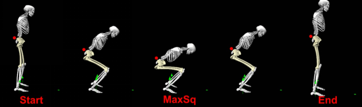
图2 下蹲序列示例。每只脚的地面反作用力(绿色箭头)由地力平台记录。下蹲试验的起始点、结束点和最低点(MaxSq)由放置在骶骨上的皮肤标记(红点)的最大和最小垂直位置确定。

图3 下蹲过程中髋臼边缘负荷(AEL)的估算。(A)使用主曲率轮廓图勾勒出每侧髋关节的髋臼缘。(B)在髋臼缘的前象限(“A”)、上象限(“S”)和后象限(“P”)上标出九个时钟点。(C)通过将髋关节反作用力(JRF)在骨盆坐标系中(黑色箭头)从髋关节中心(HJC)投影到髋臼缘上的每个时钟点(例如,绿色箭头代表11点钟方向)来估算AEL的大小。JRF与边缘的夹角定义为JRF方向与AEL方向之间的夹角(例如,黑色箭头和绿色箭头之间的夹角)。图中描绘了下蹲最低点附近的髋关节JRF,其方向向后,而AEL则投影到髋臼的后上方。(D)将“髋臼边缘平面(AEP)”(黄色)拟合到髋臼缘(紫色),以测量近似髋臼边缘与髋关节中心(HJC)(黄色线)之间的距离。(A和B改编自Song等的研究)。

图4 髋关节反作用力(JRF)的合力和前后、上下、内外分量在整个下蹲试验过程中的变化,取发育性髋关节发育不良(DDH)患者和对照组的平均值。红色/黑色阴影 = ±1 个标准差。黄色竖线表示下蹲最低点(MaxSq)的时间。*组间差异具有统计学意义。BW,体重;SD,标准差。

图5 显示了下蹲试验过程中,髋臼上部(11点至1点钟方向)和后部(8点至10点钟方向)区域的平均关节反作用力(JRF)与边缘角度(上图)和髋臼边缘负荷(AEL)(下图)。前部区域(2点至4点钟方向)的AEL值极小,且未发现组间差异,因此未显示。红色/黑色阴影表示 ±1 个标准差。黄色竖线表示最大平方和(MaxSq)的时间。蓝色阴影表示整个下蹲试验过程中的累积冲量。组间统计学显著性:*表示瞬时值,。BW,体重;SD,标准差。
Elevated loading at the posterior acetabular edge of dysplastic hips during double-legged squat
Hips with developmental dysplasia (DDH) are at a heightened risk of premature hip osteoarthritis, which is often expedited by mechanically induced articular tissue damage. A prevalent form of damage in DDH is labral tears caused by abnormal loading at the shallow acetabular edge. Although the majority of reported DDH-related labral tears occur in the antero-superior acetabulum, posterior labral tears are prevalent in individuals whose lifestyle involves frequent high hip flexion tasks such as squatting. To better understand region-specific risks for chondrolabral damage during high hip flexion, we used image-based musculoskeletal models to compare acetabular edge loading (AEL) during double-legged squat between hips with symptomatic DDH (n = 10) and healthy controls (n = 10). Compared to controls, hips with DDH had higher instantaneous posterior AEL at the lowest point of squat (2.6 vs. 1.8 ×BW, p ≤ 0.04), and higher accumulative loading across the duration of the squatting motion (2.6 vs. 1.9 ×BW*s, p ≤ 0.04). Elevated posterior AEL coincided with increased net hip extension moments and posterior joint reaction forces, and was correlated with the severity of DDH acetabular deformity. Our findings suggest that posterior AEL is elevated in hips with symptomatic DDH during double-legged squat, which may contribute to chondrolabral damage in individuals who often perform such high hip flexion tasks. Clinical evaluation of DDH should consider patient-specific anatomy and lifestyle factors when establishing diagnoses and planning personalized treatment.
文献出处:Ke Song, Cecilia Pascual-Garrido, John C Clohisy, Michael D Harris. Elevated loading at the posterior acetabular edge of dysplastic hips during double-legged squat. J Orthop Res. 2022 Sep;40(9):2147-2155. doi: 10.1002/jor.25249. Epub 2022 Jan 11.
文献4
下一代手术导航:无标记多视图六自由度手术器械位姿估计
译者 邱兴
传统计算机视觉的最前沿研究正越来越多地被应用于手术领域。计算机辅助手术中的一个特别关注点,是采用基于深度学习的纯图像6DoF位姿估计方法,取代基于标记的定位系统来实现器械追踪。然而,当前最先进的单视角位姿估计方法尚未达到手术导航所需的精度。在此背景下,我们研究了多视角设置对于实现高精度、抗遮挡的手术器械6DoF位姿估计的优势,并提出了一套应对手术室实际挑战的理想摄像系统建议。
我们的贡献主要有三方面。第一,我们提出一个多视角RGB-D视频数据集,数据来源于离体脊柱手术实验,由固定式摄像头和头戴式摄像头采集,并包含对术者、器械及患者解剖结构的丰富标注。第二,我们对三种最先进的单视角与多视角位姿估计方法进行了广泛评估,分析了相机数量与布放位置、有限的真实世界数据、以及静态、混合或全移动相机设置对位姿精度、抗遮挡鲁棒性和泛化能力的影响。第三,我们设计了一套用于无标记手术器械追踪的多摄像机系统,在最优条件下,对手术钻的平均位置误差为1.01 mm、姿态角误差为0.89°,对螺丝刀的平均位置误差为2.79 mm、姿态角误差为3.33°。我们的结果表明,无标记手术器械追踪正逐渐成为现有标记追踪系统的可行替代方案。

图1. 单视角(a)与多视角(b)及(c)下的定性结果。如(a)所示,SurfEmb 对重度截断和遮挡具有鲁棒性。对于 EpiSurfEmb,展示了五个输入视角中的三个。顶行显示输入的 RGB 图像块,底行叠加显示了估计的位姿。

图2. 深度细化步骤中的一个示例帧,其采样点大部分来自被遮挡像素(此处为术者手部)。左图展示了输入的 RGB 图像块,并高亮显示了所选像素。右图显示了预测的查询图像,其中颜色越亮表示范数越大。红点表示用于细化位姿的射线方向。

图3. 最佳单视角、双视角和五视角基线方法在 OR-X 暗光子集上的定性比较。第二行用绿色叠加了真实位姿,后续行用橙色显示估计位姿。左上角的黄色三角形表示该帧未作为位姿估计方法的输入。需要注意的是,单视角位姿估计在输入图像上具有很好的视觉重叠性,但由于深度模糊,从其他视角观察时会存在显著误差。
Next-generation surgical navigation: Marker-less multi-view 6DoF pose estimation of surgical instruments
State-of-the-art research of traditional computer vision is increasingly leveraged in the surgical domain. A particular focus in computer-assisted surgery is to replace marker-based tracking systems for instrument localization with pure image-based 6DoF pose estimation using deep-learning methods. However, state-of-the-art single-view pose estimation methods do not yet meet the accuracy required for surgical navigation. In this context, we investigate the benefits of multi-view setups for highly accurate and occlusion-robust 6DoF pose estimation of surgical instruments and derive recommendations for an ideal camera system that addresses the challenges in the operating room. Our contributions are threefold. First, we present a multi-view RGB-D video dataset of ex-vivo spine surgeries, captured with static and head-mounted cameras and including rich annotations for surgeon, instruments, and patient anatomy. Second, we perform an extensive evaluation of three state-of-the-art single-view and multi-view pose estimation methods, analyzing the impact of camera quantities and positioning, limited real-world data, and static, hybrid, or fully mobile camera setups on the pose accuracy, occlusion robustness, and generalizability. Third, we design a multi-camera system for marker-less surgical instrument tracking, achieving an average position error of 1.01mm and orientation error of 0.89° for a surgical drill, and 2.79mm and 3.33° for a screwdriver under optimal conditions. Our results demonstrate that marker-less tracking of surgical instruments is becoming a feasible alternative to existing marker-based systems.
文献出处:Hein, J., Cavalcanti, N., Suter, D., Zingg, L., Carrillo, F., Calvet, L., ... & Fürnstahl, P. (2025). Next-generation surgical navigation: Marker-less multi-view 6DoF pose estimation of surgical instruments. Medical Image Analysis, 103, 103613.
文献5
基于MRI的髋关节三维模型结合深度学习自动分割,可实现无需透视的计算机辅助髋臼周围截骨术规划
译者 徐子茵
引言: 髋关节发育不良(developmental dysplasia of the hip,DDH)和股骨髋臼撞击综合征(femoroacetabular impingement,FAI)均为复杂的三维髋关节病变,可导致年轻患者出现髋痛和骨关节炎。基于三维MRI的模型可用于无辐射的计算机辅助手术规划。由于手动分割耗时较长,因此更倾向于采用MRI三维模型的自动分割方法。
本研究旨在探讨:(1)基于自动MRI分割的三维模型与手动CT分割的三维模型在股骨头覆盖率方面的差异;(2)二者在股骨头覆盖率方面的相关性;以及(3)该方法在有症状髋关节疾病患者术前规划中的可行性。
方法: 本研究为经机构审查委员会批准的比较性回顾性研究,共纳入31髋,来自26例有症状的髋关节发育不良或FAI患者。所有患者均接受髋关节三维MRI序列和CT扫描。术前MRI包括髋关节轴斜位T1 VIBE序列,体素大小为0.8 mm³各向同性。研究者分别对MRI和CT图像进行手动分割,并使用深度学习方法对基于MRI的三维模型进行自动分割。
结果: (1)基于MRI的三维髋关节模型中,自动分割与手动分割之间的差异小于1 mm,其中近端股骨为0.2 ± 0.1 mm,髋臼为0.3 ± 0.5 mm。近端股骨和髋臼的Dice系数分别为98%和97%。(2)自动MRI三维模型与手动CT三维模型之间,总股骨头覆盖率的相关性优异且具有统计学意义,r = 0.975,p < 0.001。自动MRI三维模型与手动MRI三维模型之间,总股骨头覆盖率的相关性同样优异,r = 0.979,p < 0.001。(3)对于所有髋关节发育不良或髋臼后倾患者,基于该方法进行髋臼周围截骨术的术前规划和模拟均可行,可行率为100%。
结论: 对于育龄期髋关节疾病患者,采用深度学习对MRI三维模型进行自动分割,其准确性与基于CT的三维模型相当。该方法可为DDH患者提供无需透视、个体化的髋臼周围截骨术前模拟和手术规划。
MRI-based 3D models of the hip joint enables radiation-free computer-assisted planning of periacetabular osteotomy for treatment of hip dysplasia using deep learning for automatic segmentation
Introduction: Both Hip Dysplasia(DDH) and Femoro-acetabular-Impingement(FAI) are complex three-dimensional hip pathologies causing hip pain and osteoarthritis in young patients. 3D-MRI-based models were used for radiation-free computer-assisted surgical planning. Automatic segmentation of MRI-based 3D-models are preferred because manual segmentation is time-consuming.
To investigate(1) the difference and(2) the correlation for femoral head coverage(FHC) between automatic MR-based and manual CT-based 3D-models and (3) feasibility of preoperative planning in symptomatic patients with hip diseases.
Methods: We performed an IRB-approved comparative, retrospective study of 31 hips(26 symptomatic patients with hip dysplasia or FAI). 3D MRI sequences and CT scans of the hip were acquired. Preoperative MRI included axial-oblique T1 VIBE sequence(0.8 mm³ isovoxel) of the hip joint. Manual segmentation of MRI and CT scans were performed. Automatic segmentation of MRI-based 3D-models was performed using deep learning.
Results: (1)The difference between automatic and manual segmentation of MRI-based 3D hip joint models was below 1 mm(proximal femur 0.2 ± 0.1 mm and acetabulum 0.3 ± 0.5 mm). Dice coefficients of the proximal femur and the acetabulum were 98 % and 97 %, respectively. (2)The correlation for total FHC was excellent and significant(r = 0.975, p < 0.001) between automatic MRI-based and manual CT-based 3D-models. Correlation for total FHC (r = 0.979, p < 0.001) between automatic and manual MR-based 3D models was excellent. (3)Preoperative planning and simulation of periacetabular osteotomy was feasible in all patients(100 %) with hip dysplasia or acetabular retroversion.
Conclusions: Automatic segmentation of MRI-based 3D-models using deep learning is as accurate as CT-based 3D-models for patients with hip diseases of childbearing age. This allows radiation-free and patient-specific preoperative simulation and surgical planning of periacetabular osteotomy for patients with DDH.
文献来源:Zeng G, Schmaranzer F, Degonda C, Gerber N, Gerber K, Tannast M, Burger J, Siebenrock KA, Zheng G, Lerch TD. MRI-based 3D models of the hip joint enables radiation-free computer-assisted planning of periacetabular osteotomy for treatment of hip dysplasia using deep learning for automatic segmentation. Eur J Radiol Open. 2020 Dec 18;8:100303. doi: 10.1016/j.ejro.2020.100303. PMID: 33364259; PMCID: PMC7753932.
来源:304关节学术
作者:304关节团队
声明:本文内容及图片均为转载内容,如涉及版权问题请相关权利人及时与我们联系,我们会立即处理配合采取保护措施,以保障双方利益。
为什么要投稿?是为了记录自己的医学之路!是为了与更多的骨科同道交流分享!是为了让更多的人看到而受益!让传播知识成为一种习惯,是“玖玖骨科”让你投稿的理由!


热门跟贴